CT Scan CVJ (CT cranio-vertebral junction)
4.6 (5 reviews)A CT scan of the cranio-vertebral junction to evaluate bone alignment and structural abnormalities.
Test Details
Also Known As
CT CV Junction, CT Craniovertebral Junction, CT Atlanto-Occipital Joint
Sample Type
CT imaging of cranio-vertebral junction, no contrast dye required
Gender
Male, Female
Age Group
All Age
Categories
CT
Test Code
Radiology
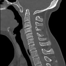
CT Scan CVJ is a specialized CT scan used to evaluate the cranio-vertebral junction, where the skull base meets the upper cervical spine. It provides detailed assessment of the atlas (C1), axis (C2), occipital bone, and surrounding bony structures. This scan is commonly used to diagnose congenital anomalies, trauma, fractures, atlanto-axial instability, basilar invagination, and degenerative changes. CT CVJ offers high-resolution images that are crucial for accurate diagnosis and surgical planning, especially in trauma and neurosurgical cases. The procedure is quick, non-invasive, and involves controlled radiation exposure when clinically indicated.
Recommended Tests
₹69.00 ₹200.00
₹69.00 ₹200.00
₹399.00 ₹800.00
₹299.00 ₹400.00
₹299.00 ₹600.00
₹399.00 ₹800.00
₹79.00 ₹200.00

₹1350.00 ₹2500.00
₹699.00 ₹1500.00