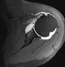
MRI Arthrogram
4.6 (5 reviews)A specialized MRI scan with joint contrast to detect subtle joint and cartilage injuries.
Test Details
Also Known As
MR Arthrography, MRI Joint Arthrogram, Contrast MRI Arthrogram
Sample Type
MRI imaging with intra-articular contrast injection
Gender
Male, Female
Age Group
All Age
Categories
MRI
Test Code
Radiology
MRI Arthrogram is an advanced imaging procedure used to evaluate joints in detail by injecting contrast material directly into the joint space before MRI scanning. This technique enhances visualization of joint structures such as cartilage, ligaments, labrum, tendons, and joint capsule. It is especially useful for diagnosing labral tears, ligament injuries, cartilage defects, and post-surgical complications that may not be clearly visible on routine MRI. MRI Arthrogram is commonly performed for shoulder, hip, wrist, and ankle joints. The test provides high diagnostic accuracy, helping doctors plan appropriate treatment or surgical intervention.
Recommended Tests
₹69.00 ₹200.00
₹69.00 ₹200.00

₹299.00 ₹800.00
₹179.00 ₹400.00
₹250.00 ₹600.00
₹499.00 ₹800.00

₹79.00 ₹200.00

₹1350.00 ₹2500.00
₹699.00 ₹1500.00