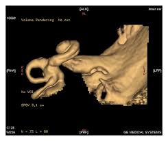
MRI Brain with Inner Ear (3D Cochlea)
4.6 (5 reviews)An advanced MRI scan to evaluate the inner ear, cochlea, and related brain structures in detail.
₹3999.00 ₹14000.00
Test Details
Also Known As
MRI Inner Ear, 3D Cochlea MRI, MRI Brain with IAC, Inner Ear MRI Scan
Sample Type
MRI imaging, contrast dye if advised
Gender
Male, Female
Age Group
All Age
Categories
MRI
Test Code
Radiology
An MRI of the brain with a focus on the inner ear, particularly the cochlea in three dimensions (3D), is a specialized imaging technique that provides detailed views of the inner ear structures, including the cochlea, auditory nerve, and surrounding soft tissues. This scan helps doctors evaluate the inner ear for conditions like malformations, tumors, or infections. 3D images can be reconstructed to show the cochlea and other inner ear structures in greater detail, making it useful for diagnosing and managing hearing loss and other ear-related issues.
-
Purpose:
- To assess the cochlea for normal shape and any abnormalities.
- To evaluate the hearing nerve and surrounding soft tissues.
- To diagnose conditions like infections, tumors, or hemorrhage in the inner ear.
- To help identify potential causes of hearing loss or tinnitus.
Recommended Tests

₹69.00 ₹200.00
₹69.00 ₹200.00

₹399.00 ₹800.00
₹299.00 ₹400.00
₹299.00 ₹600.00

₹399.00 ₹800.00

₹79.00 ₹200.00

₹1350.00 ₹2500.00
₹699.00 ₹1500.00

₹550.00 ₹1250.00
Frequently Asked Questions
It is used to evaluate hearing loss, vertigo, tinnitus, and inner ear abnormalities.
Yes, it is safe and does not involve radiation.
Contrast may be used depending on the suspected condition.
Fasting is usually not required unless contrast is planned.
The scan typically takes about 40 to 60 minutes.
It is recommended for patients with unexplained hearing loss, balance issues, or suspected inner ear disorders.